
Unlike people, dogs have three eyelids, two of them are covered with hair and we see them with the naked eye (lower and upper), and the third is “hidden” under the eyelids and is not visible under normal conditions.
Sometimes this third eyelid can be made visible by appearing from the tear area as a thin, whitish-pinkish membrane that will cover part of the surface of the eye. Let’s see what are the main problems that this third eyelid can cause and what solution they have.
Index of contents
- 1 What is the third eyelid of dogs?
- 2 What is the third eyelid for dogs?
- 3 When does the third eyelid become visible?
- 4 What are the main alterations of the third eyelid in dogs?
- 4.1 What is the protrusion of the third eyelid?
- 4.2 What is Harder’s gland prolapse?
- 4.2.1 How is cherry eye treated?
- 4.3 Neoplasms of the third eyelid
What is the third eyelid of dogs?
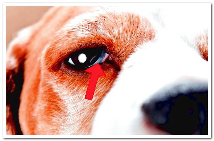
Dogs, in addition to the two visible eyelids (upper and lower), they have a third eyelid called the nictitating membrane.
This additional eyelid, It is a thin membrane of connective tissue that is located in the medial canthus of the eye, hidden under the eyelids.. It is not covered with hair like the other two eyelids, nor does it have musculature, but it does have a lacrimal gland called the Harder’s gland.
Under normal conditions, the dog’s third eyelid is not visible, but it can exteriorize under certain circumstances and become visible as a whitish-pinkish membrane that will cover part of the surface of the eye.
What is the third eyelid for dogs?
The third eyelid or nictitating membrane has two main functions in the dog:
- Protective function: When a foreign particle enters the dog’s eye, the eyeball retracts into the orbit and the third eyelid appears to cover part of its surface; This membrane can help remove foreign bodies due to a drag effect, as if it were a windshield wiper, being a reflex mechanism to avoid eye injuries. It can also be exteriorized in case there are alterations in the eye, such as conjunctivitis or ulcers, acting as a protective layer until the injury resolves.
- Moisturizing function: The third eyelid has a lacrimal gland (Harder’s gland) that is responsible for around 40% of the total tear production, therefore, it is essential to maintain a correct degree of hydration in the eye.
When does the third eyelid become visible?
As we have seen, the nictitating membrane It is exteriorized covering the surface of the eye as a defense mechanism against irritations or alterations that can cause damage to the eyes.
So if we look at the third eyelid in our dog, it is very likely that he has a foreign particle in his eye or an already established alteration / injury to the eye (conjunctivitis, ulcer, etc.).
It is also frequent to observe an externalization of the third eyelid in dogs that have “discomfort” or some general pathology (infectious process, gastroenteritis, fever, etc.)
It is because of that the third eyelid can serve as an “alarm” indicative that our dog may be suffering an ocular alteration or his general health.
What are the main alterations of the third eyelid in dogs?
Mainly we can describe three processes that affect the nictitating membrane in dogs:
- Protrusion of the third eyelid
- Prolapse of the Harder’s gland or cherry eye
- Neoplasms of the third eyelid
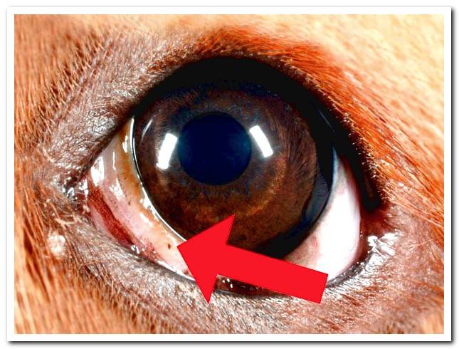
What is the protrusion of the third eyelid?
The nictitating membrane in normal conditions is not visible, and it is exteriorized as a defense mechanism against eye injuries or in situations of general diseases such as feverish, gastrointestinal processes, etc.
If we observe that a whitish pinkish membrane appears in the eye / s of our dog from the medial canthus of the eye (lacrimal area) covering part of the ocular surface, and up to 6 hours pass without this membrane returning to its place and ceasing to be visible, we should consult your veterinaryn, since, although it is not an emergency situation, it may be indicative that there is an ocular alteration or even that our pet has some underlying general disease.
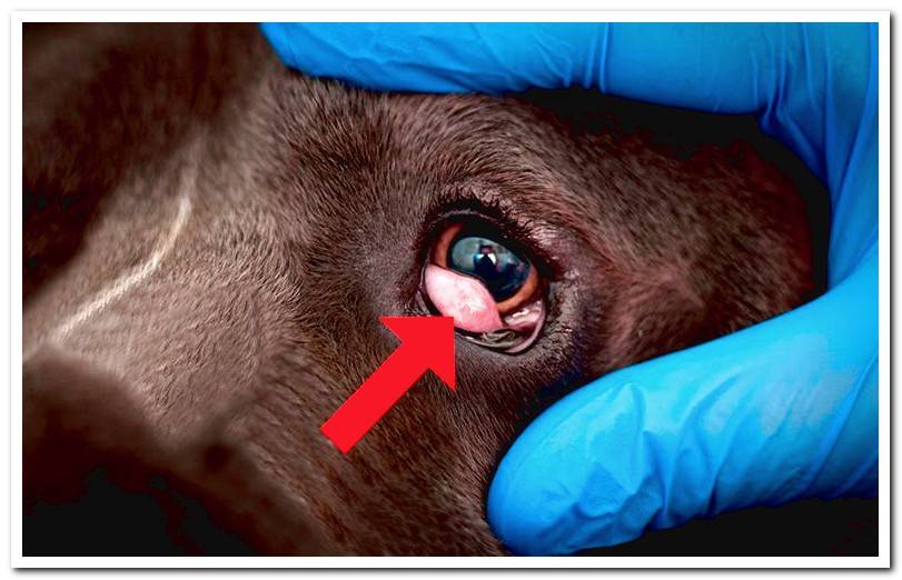
What is Harder’s gland prolapse?
The nictitating membrane has at its base a tear-secreting gland (Harder’s gland) that is fixed in position by bands of connective tissue.
If this fixation is weak, a prolapse of the gland can occur to the outside, appearing a pink ball (mass) in the area near the tear of the eye.
This type of alteration is known as cherry eye, and it is a process that is observed quite frequently in the veterinary practice. Although it can occur in any dog, there are certain breeds that are predisposed to suffer this alteration: Bulldog, Boxer, Hound, Beagle, Pekingese, Chihuahua, Cocker Spaniel, among others.
How is cherry eye treated?
This ocular alteration it must be treated by a veterinaryn as soon as possible, since it can cause alterations in the eye such as keratoconjunctivitis sicca, when the production of tears was compromised.
Initially, a topical anti-inflammatory treatment (eye drops with corticosteroids) is established to decrease the volume of the affected area. Subsequently, under anesthesia, the prolapse is reduced (the gland is relocated in its place) and to the fixation of the gland by means of sutures to avoid that it prolapses again.
Once the intervention is completed, antibiotic and anti-inflammatory therapy should be established by administering eye drops and preventing the animal f
rom making efforts that can cause tension in the suture area. It is essential to place a Elizabethan necklace to prevent the dog from scratching the area until its full recovery.
Neoplasms of the third eyelid
Although nictitating membrane tumors are not very frequent, they can occur especially in large breed dogs of advanced age. The most common tumor is melanoma and tends to be malignant with a probability of frequent metastasis. Tumors can also affect Harder’s gland called adenocarcinomas.
The diagnosis of tumor processes is carried out by means of fine-needle aspiration to analyze the integrating cells and its treatment consists of complete surgical excision of the nictitating membrane. Adjuvant techniques such as cryotherapy, laser or radiotherapy.
As we can see, the third eyelid has a very important role in the eye health of our pet So if we observe any of these alterations in our dog, we should not let them pass, and we must go to your vet as soon as possible, since early treatment is the basis to avoid serious eye damage.